I - V 9 a.m. - 8 p.m.
I - V 2 p.m. - 3 p.m. concilium of doctors
VI - VII 9 a.m. - 5 p.m.
When people hear the word “cancer”, they often think it’s a death sentence. And indeed, cancer is one of the most insidious and difficult diseases not only in human medicine, but also in veterinary medicine, especially if it is detected too late, i.e. at an advanced stage. Early diagnosis is particularly important in oncology.
Siaurio Šnauceris has more than 30 years of experience in veterinary oncological diseases. We were among the first in the Baltic States to offer chemotherapy treatment, and were the first to successfully apply a breakthrough non-surgical method of treating mastocytoma with special drugs. We are constantly looking for the latest treatment methods and introducing them at our clinics.
The term “cancer” refers to diseases characterised by uncontrolled cell growth/division, resulting in the formation of a mass of tissue (a tumour). That is, with the exception of blood cancer, which does not cause visible tumours. Cancer cells grow faster than normal cells and can spread anywhere in the body; if left untreated, they can continue to spread.
Tumours can be benign or malignant (cancerous). Benign tumours grow without penetrating or spreading to adjacent and distant organs. However, as benign tumours grow, they can press on adjacent organs and cause health problems. In most cases, they are successfully removed by surgery, and regrowth is rare. The cells of a benign tumour are not very different from the surrounding cells – they are just unusually arranged.
The cells of malignant tumours are a lot different from the cells of the healthy tissue from which they developed – they are immature and not fully differentiated. The nature of the tumour growth is different – the cells penetrate deeper and deeper into the tissue of the organ from which it developed, and grow into adjacent organs. The structure of the organs is damaged, their activity is impaired, and serious health disorders appear. In addition, malignant tumour cells have the ability to break away from the tumour and spread through lymph or blood to regional lymph nodes and other organs of the body, including ones that are often distant from the primary tumour. Once they reach a new site, these cells “take root” there and multiply. New, or secondary, tumours then form. This is called metastasis. Metastasis complicates the course of the disease significantly.
There is no strict boundary between benign and malignant tumours – some benign tumours can become malignant.
There is no single answer as to why some pets develop cancer. Approximately one in four dogs get cancer; incidentally, this rate is similar for cats. Cancer is one of the most common causes of death in older animals.
The tissues of an animal’s body, like a person’s, are made up of the smallest fundamental unit of structure – cells. The cells of one organ differ from the cells of another organ in their appearance and function, but their division and reproduction processes are similar. Healthy cells divide in an orderly manner because the body controls the division. Sometimes, when exposed to harmful factors (radiation, chemicals, hormones or infections), an organ’s cells are damaged over time to the point that they begin to defy the body’s control – they divide incessantly. An accumulation of altered cells is formed – a tumour.
All mammals have immune defence mechanisms to prevent or repair cell damage. However, these defence mechanisms are not perfect. Some have defects in their defences, so cancer is more common than expected. Those defects can be congenital – for example, some purebred dogs have a hereditary predisposition to certain cancers. Other defects appear over time. In some cases, the defence mechanisms simply cannot cope with an excessive amount of external environmental factors.
It is incredibly difficult to list specific symptoms, since each type of cancer is different, but one of the most obvious signs of cancer is a lump that gets bigger. The neoplasm may ulcerate, bleed or cause other physical discomfort. However, cancer can develop in any cell (even in a blood cell, in the case of blood cancer) and in any part of the body, and internal organs are no exception. In this case, there are no external signs, and symptoms do not appear immediately. Still, there are some tell-tale signs that signal a possible cancer:
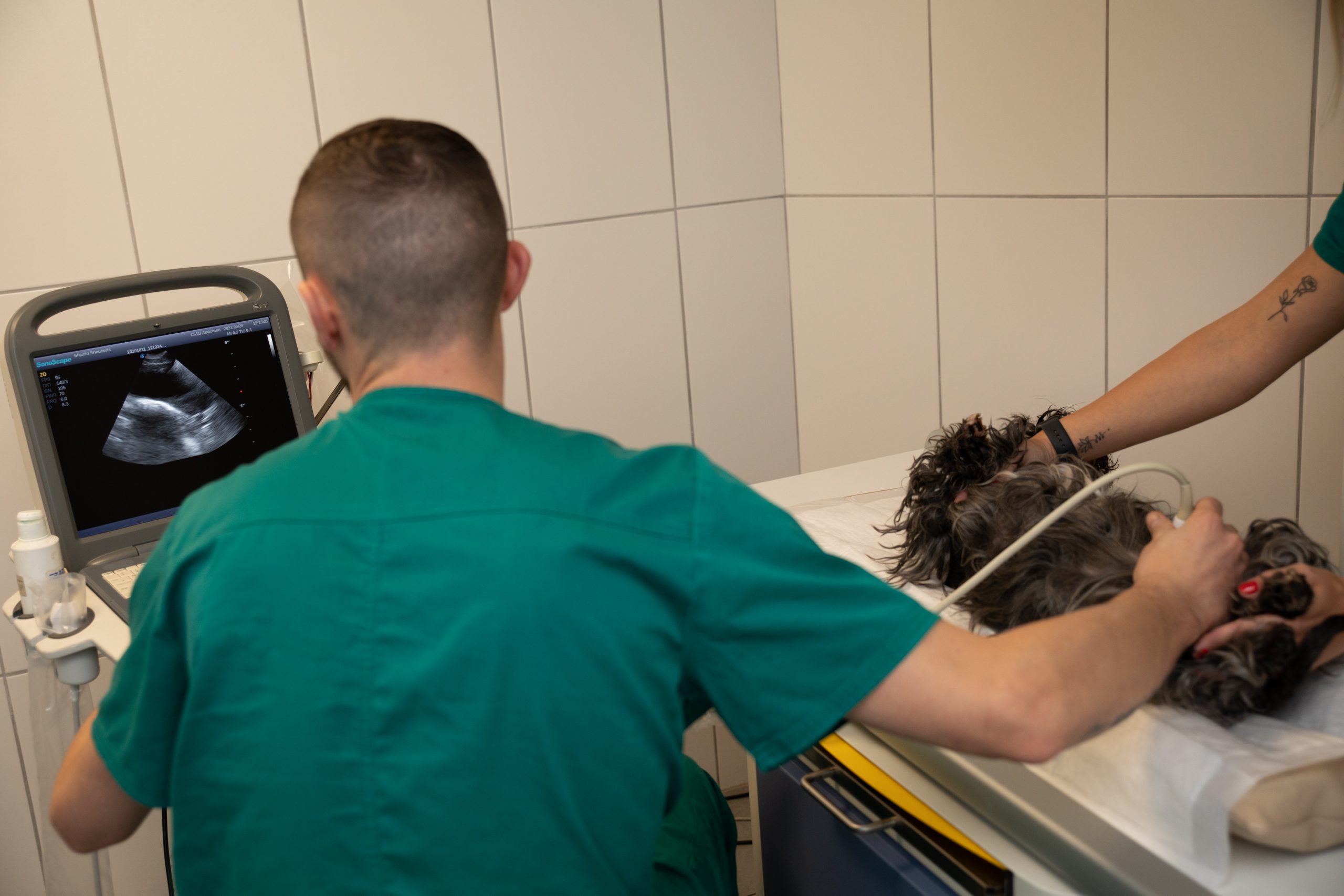
A veterinarian may suspect that your pet has cancer based on clinical signs. Subsequently, the following tests may be performed as needed:
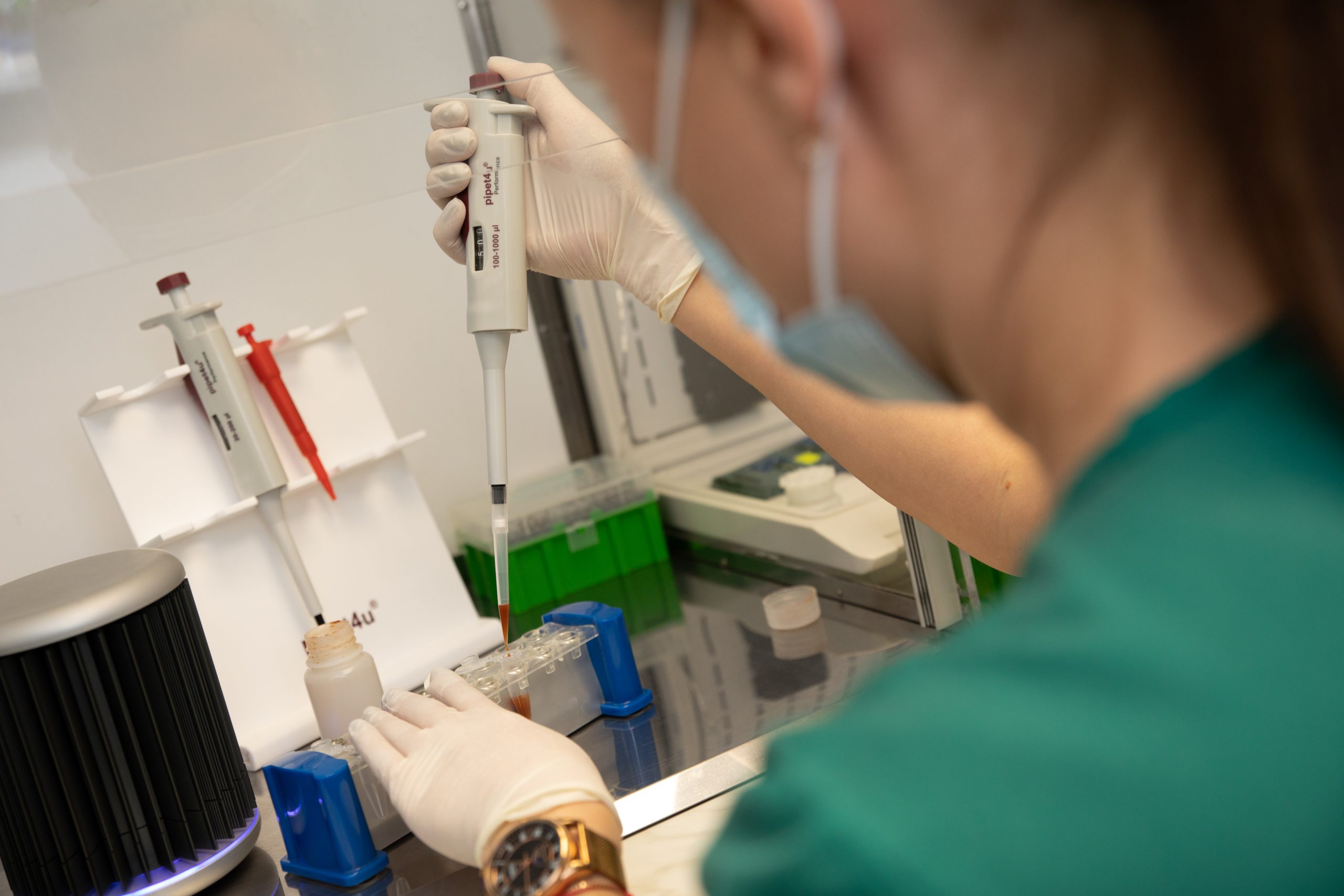
At Siaurio Šnauceris, we perform cytological and histopathological (LABOKLIN) examinations to accurately determine the type of cancer. Some tumours can be accurately diagnosed by cytology tests, but a tissue biopsy is usually required to make an accurate and reliable diagnosis. In that case, we send the sample to the specialised LABOKLIN laboratory in Germany for evaluation by a veterinary pathologist. Histopathology involves examining tissues under a microscope. The histopathology report usually indicates whether the tumour is benign or malignant.
At our clinic, we do blood tests to screen dogs for prostate cancer with the CPSE biomarker.
Most malignant tumours originate from epithelial cells. They are called carcinoma. Another type of malignant tumours is sarcomas, which occur not in epithelial cells, but in cartilage, bone, fat, muscle cells or vascular tissues.
The course of the disease and its treatment are closely related to the type of malignant tumour, the degree of differentiation of tumour cells and the spread of the cancer.
SURGICAL – RADICAL REMOVAL OF THE TUMOUR
The most common treatment for cancer is surgical removal of the tumour. This method of treatment is most effective in cases where the tumour has clear boundaries and there is no metastasis to other organs or lymph nodes. The surgical method cannot be used for a non-tumour types of cancer (e.g. leukaemia). In these cases, chemotherapy is used.
CHEMOTHERAPY
Chemotherapy is the treatment of cancer with special drugs that inhibit the essential property of the cancer cells – their rapid, uncontrolled division. When cancer cells lose their ability to multiply, they die, and the tumour stops growing. These drugs, after entering the blood stream, are carried to the tissues of the body and directly affect the tumour cells. Rapidly proliferating cells are most susceptible to the effects of these drugs. Because cancer cells multiply and grow much faster than normal, healthy cells in the body, they are the first and most affected by chemotherapy. However, healthy cells are also inevitably damaged, just to a much lesser extent, so they are able to recover. It is important to mention that animals tolerate chemotherapy much better than humans, and side effects such as vomiting, hair loss and so on are very mild or do not occur at all.
Chemotherapy for animals is done at the veterinary clinic, during which special drugs are delivered into the vein with an IV. The session lasts 30-60 minutes. After the procedure, the animal is left in the clinic’s day hospital under the supervision of veterinarians, who monitor its condition. When an animal is getting chemotherapy treatment, we periodically perform arterial blood gas tests and monitor the biochemical parameters of the internal organs. The veterinarian can tell you how many chemotherapy sessions will be needed to overcome the cancer after evaluating the results of the cytological or histopathological examination. Only by knowing how advanced the disease is can the treatment protocol that will give the best outcome be selected (number of sessions, frequency).
Chemotherapy is usually used in combination with surgical treatment to destroy macroscopically invisible cancer cells and prevent metastasis, or to stop the proliferation of remaining cancer tissue that cannot be completely removed (without clear boundaries).
We were one of the first veterinary clinics in Lithuania to start using chemotherapy, so we have extensive experience in this treatment. Siaurio Šnauceris has been using chemotherapy for many years to save pets’ lives.
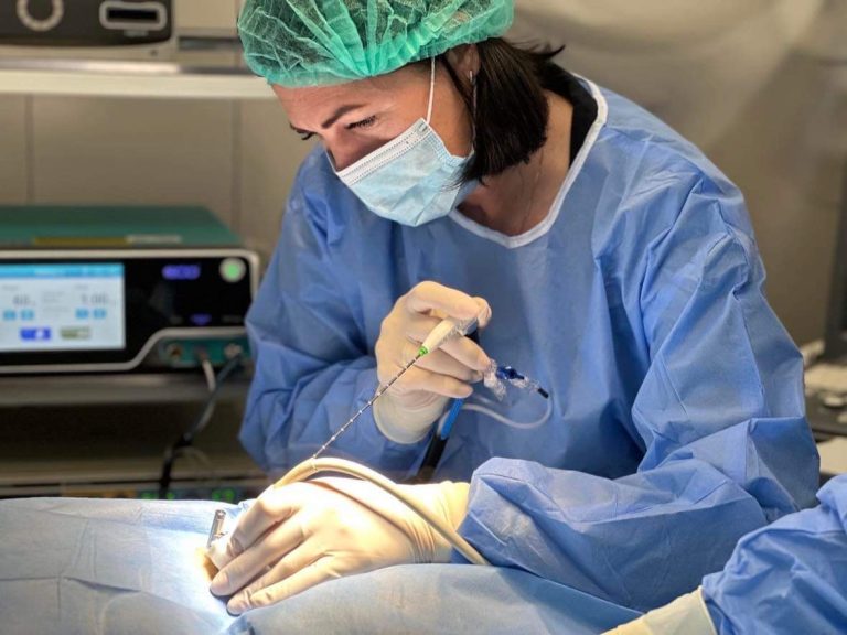
We are thrilled to present a new-generation microwave ablation system for solid tumours, which is also used in veterinary medicine, and which allows us to perform minimally invasive surgeries. Ablation therapy is a minimally invasive procedure that destroys malignant and benign solid tumours of the organs, skin and so on through a needle puncture. During the procedure, the tumour is destroyed by inserting a special needle or needles into it and exposing it to microwave energy.
This modern method, which is still being used in human medicine now, has finally made its way to veterinary medicine as well, and our clinic is one of the few in Lithuania that can offer this treatment.
Siaurio Šnauceris has more than 30 years of experience in veterinary oncological diseases, and we were one of the first to offer chemotherapy treatment. Now, we can help your pets with this modern system as well.
Mastocytoma is a relatively common skin and subcutaneous cancer that, if left untreated, can spread to other organs – the lymph nodes, spleen, liver or bone marrow. Mastocytoma is diagnosed by cytological sampling of the neoplasm; a biopsy is not necessary.
There are several methods of treating mastocytoma, which the doctor will recommend individually for each patient:
We are extremely proud to be the first clinic in Lithuania and the Baltic States to successfully apply the newest medicinal method of treating mastocytoma.
Chemotherapy, 1 procedure (not including blood tests) 120 – 250
Microwave ablation 100 – 300
Treatment of mastocytoma with special drugs from 450
Removal of the mammary gland 90 – 160
Total removal of the one side of the mammary glands 150 -250
Removal of externally visible formations 60 – 210
Preliminary prices are indicated. The specific price is given after evaluating the case of each patient individually
This story is really special and unique not only in Lithuania, but also in the Baltic States.
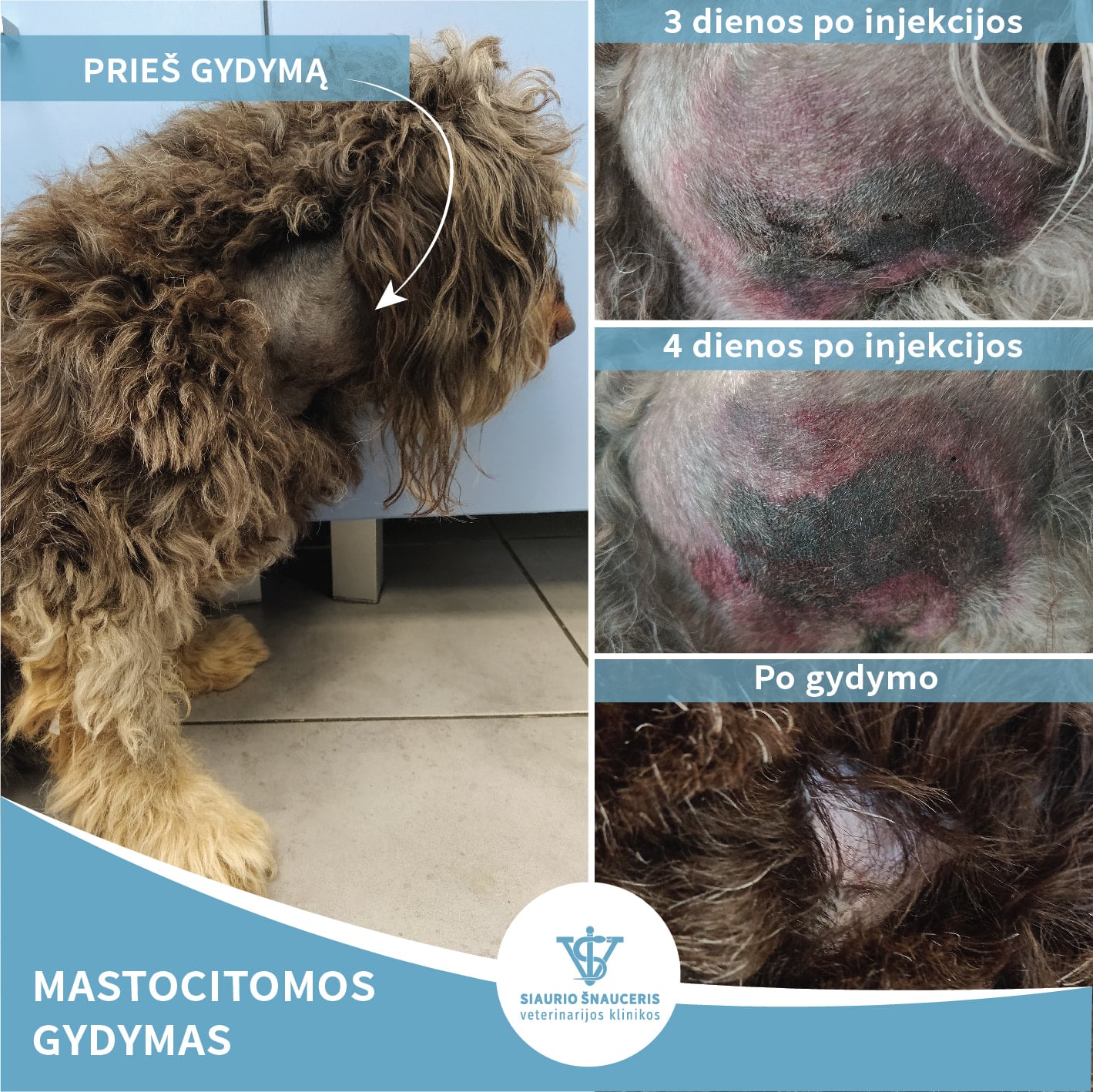
Rikis, an 8-year-old crossbreed, came to the clinic because of a tumour on the right side of his neck. A cytology specimen was taken from the neoplasm, and the same day the diagnosis was confirmed in the clinic laboratory – mastocytoma.
Mastocytoma is a tumour of mast cells. This type of tumour is highly invasive, metastatic and the most common round cell tumour in dogs.
Therefore, we are extremely happy and proud to be the first in Lithuania and the Baltic States to use this breakthrough injectable solution from Virbac to treat mastocytoma to successfully treat this patient. Injected into the neoplasm, the drug destroys the tumour cells without damaging healthy tissues. This treatment can be used in the diagnosis of mastocytoma, sarcoma or melanoma.
Rikis needed one injection and three months to fully recover. We are delighted with the outcome!
Kingas, a 12-year-old Labrador, came to the clinic because of the sudden onset of a large (approximately 8.5 cm) abdominal swelling. A cytology specimen was taken from the neoplasm, and the diagnosis was confirmed in the clinic laboratory – mastocytoma. As we mentioned earlier, mastocytoma is a tumour of mast cells.
We used the breakthrough injectable solution from Virbac to treat Kingas. Injected into the neoplasm, the drug destroys the tumour cells without damaging healthy tissues. This treatment can be used in the diagnosis of mastocytoma, sarcoma or melanoma.
In the photo, you can see how Kingas’s abdomen looked 55 days after the treatment – the neoplasm had disappeared. However, during regular follow-up examinations, it was detected that due to its size, the neoplasm had not been completely eliminated. A second dose of the drug was therefore injected, which is successfully destroying the tumour at this very moment.
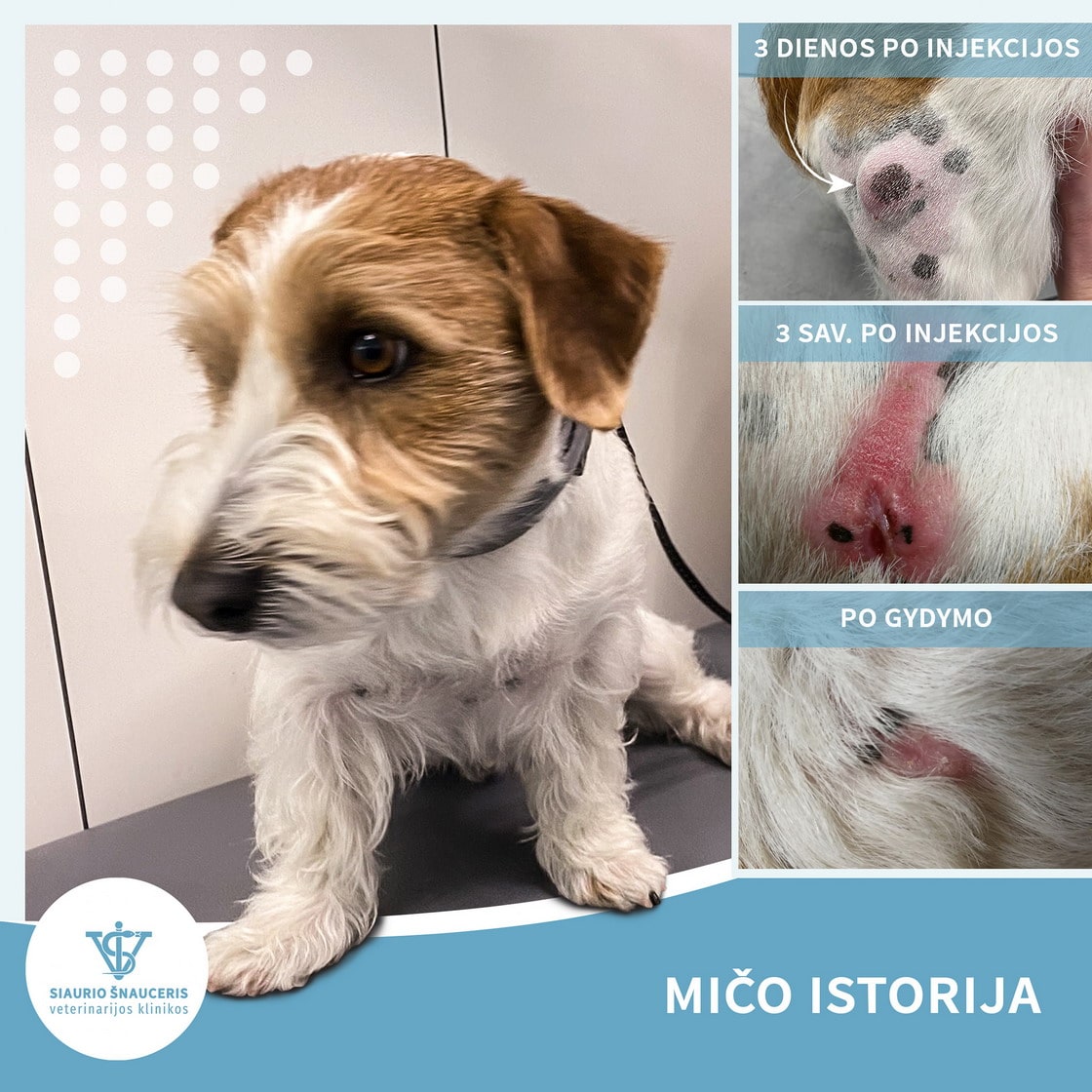
We want to share the story of Mičas, a 3 1/2-year-old Jack Russell Terrier.
This little guy came to see us because of a suspicious growth on his hind leg. After performing a cytology test, the diagnosis was mastocytoma (round cell tumour). The treatment started shortly after with a special injection directly into the neoplasm, which, as you can see from the photos, did its job perfectly. Now, 30 days after that single injection, the tumour has been completely destroyed and Mičas is fully recovered!